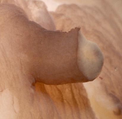

Figure. ventrolateral view of Mg. microlucens, paratype, swimming in an aquarium after being sucked into a cold, sea-water pipe from about 925 m depth. The white spot in front of the eye is the stub of a tentacle; both tentacles were lost during capture. Photograph by Michael Darden.
- Arms
- Largest arm suckers tend to be located mid-arm but pattern highly variable. More information on relative sucker sizes can be found here.
 Click on an image to view larger version & data in a new window

Figure. Arm sucker arrangement in Mg. microlucens. Left - Oral view of selected arm suckers, paratype, 140 mm ML (estmate). All suckers maintain relative sizes. White numbers indicate sucker number counted from arm base. Sucker measurements can be found below. Photographs by R. Young. Right - Oral-oblique view of the arms and suckers of the live squid, paratype, 135 mm ML, showing mid-arm suckers on arm II that are much larger than those found mid-arm on arm IV. Color has been altered slightly to improve contrast. Photographed in an aquarium by Jan War .
- Largest arm suckers tend to be located mid-arm but pattern highly variable. More information on relative sucker sizes can be found here.
- Tentacles
- Club length more than 80% of tentacle length.
- Club suckers numerous, extremely small (ca. 0.05 mm); suckers lack enlarged lateral pegs on their outer rings and have smooth inner rings.
- Base of club long, tapers gradually.
- Protective membranes present but very low (height comparable to that of individual suckers); trabeculae cannot be detected.
- Tentacular stalk not completely surrounded by club even at tip.
 Click on an image to view larger version & data in a new window
Figure. Oral view of the base of the tentacular club of Mg. microlucens, 77 mm ML. Top - Photograph of club base stained with methylene blue stain. Bottom - Artistic interpretation of the same club. Sucker-bearing portion of club in green. Illustration by R. Young.
- Club length more than 80% of tentacle length.
- Head
- Olfactory organ a short broad papilla with reduced head.
- Funnel
- Funnel pocket between bridles absent.
- Funnel pocket between bridles absent.
- Fins
- Fin length approximately equal to fin width.
- Fin length approximately equal to fin width.
- Tubercules
- Skin tubercules absent.
- Skin tubercules absent.
- Pigmentation
- Arms with dense chromatophores aborally, epithelial pigmentation orally (?).
- Head, funnel, mantle and fins with dense chromatophores.
- Most pigment beyond oral region in chromatophores.
- Measurements (from Young, et al., 2008)
Measurements in mm. ND = no data.Specimen Holotype
NMNH 1111879
Paratype
NMNH
11118800Paratype
SBMNH
422886 Teuthis 23
Paratype
SBMNH
422887
HM 1996
Paratype
SBMNH
422888
HM HawaiiParatype
SBMNH
422889
Keahole Pt.Paratype
SBMNH
422890
HM HawaiiSex Immature female Unknown Unknown Immature female Unknown- head only Immature female Mature male Mantle length
79
37 77 138 ND 135 215 Tail length 14 8.5 ND ND ND ND ND Mantle width 21 9 ND ND ND 25 ND Fin length 54 22 54 ND ND 90 ND Fin width 57 25 68 ND ND 115 ND Head width 22 9 ND ND ND ND ND Head length 16* 10 ND ND ND 34 ND Eye diameter 16** ND ND ND ND ND ND Arm I, length 34 11 44+ 87 90+ 105 125 Arm II, length 49 17 56 105+ 139 126 145 Arm III, length 36 10 43 94 111 105 137 Arm IV, length 79 35 92 110+ 174 174 ND Tentacle length ND 46 158 ND ND ND ND Club length ND 30 130 ? ND ND ND ND
*Measured laterally from olfactory organ to tentacle.
**Calculated from lens diameter (= 6 mm).
- Sucker diameter measurements from a single squid (see above)
Measurements in mm. Photographs of these suckers appear above on this page.Sucker number 2 3
13 14 15 25 26 27 34
35 43
45 50
Arm I 1.9
2.1
2.0
1.8
1.3
Arm II 2.2
2.4
3.3
3.1
2.5
1.9
Arm III 1.9 2.0
2.2
1.9
1.5
Arm IV 1.9
2.0
1.8
1.6
1.3
Comments
Not only are the photophores small (ca. 1/3 the diameter of the large integumental photophores of Mastigoteuthis spp. , but they are very difficult to recognize as photophores. The photophores lack the readily visible white reflectors seen in the Mastigoteuthis spp.; the whitish "lens" that distinguishes them as photophores are often not seen at all. Their position immediately beneath the chromatophore layer is their most apparent characteristic. In the photograph below the chromatophore layer was lost from portions of the skin shown on either side of an intact chromatophore layer. Most of the dark structures (arrows) are photophores. Where the covering of chromatophores is intact, the photophores become difficult to detect.


 Go to quick links
Go to quick search
Go to navigation for this section of the ToL site
Go to detailed links for the ToL site