Histioteuthis meleagroteuthis: Description continued
Richard E. Young and Michael Vecchione
- Arms
- Length100-150% of ML; subequal except for mature males.
- Arm suckers with 6-20 low, rounded or square teeth on distal and lateral margins (drawing at right). Basal suckers with fewer, broader teeth.

Click on an image to view larger version & data in a new window

Figure. Oral view of sucker from mid-arm II of H. meleagroteuthis, 39 mm ML, 12° 59' N, 32° 49' W. Drawing from Voss, 1969 (Fig. 26g).
- Small tubercles, fused basally, on proximal half of arms I-III along aboral midline.

Click on an image to view larger version & data in a new window

Figure. Dorsal view of head and arms, lateral view of mantle of H. meleagroteuthis, 26 mm ML, central North Atlantic. Photographed aboard the R/V G. O. SARS, Mar-Eco cruise, by R. Young.
- Tentacles
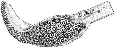
- Suckers of manus in 6-7 irregular series.
- Median suckers of the manus only slightly enlarged.
- Enlarged suckers of club manus with 25-30 short, pointed teeth around entire margin but usually longer on distal margin.

Click on an image to view larger version & data in a new window
xx

Click on an image to view larger version & data in a new window
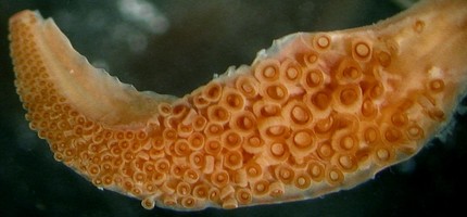
Figure. Oral view of club of H. meleagroteuthis. Top - Neotype, 48 mm ML. Drawing from Voss, 1969 (Fig. 26b). Bottom - Squid from the cental North Atlantic. Photograph by R. Young.
- Web and buccal crown
- Inner web between arms I-III 10-18% of longest arm; outer web absent.
- Buccal crown with 7 supports.

Click on an image to view larger version & data in a new window
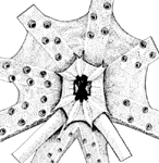
Figure. Oral view of buccal crown of H. meleagroteuthis, neotype, 48 mm ML. Drawing from Voss, 1969 (Fig. 26c).
- Funnel
- Funnel organ with dorsal pad unsculptured.

Click on an image to view larger version & data in a new window

Figure. Ventral view of dorsal pad of funnel organ of H. meleagroteuthis, neotype, 48 mm ML. Drawing from Voss, 1969 (Fig. 26e).
- Mantle
- Small tubercules form serrate ridge on middorsal line on anterior 1/2-2/3 of mantle beneath epithelium (see photograph under "Arms").
- Photophores
- Compound photophores of uniform small size, densely set, on anterior 3/4 of ventral mantle.

Click on an image to view larger version & data in a new window
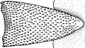
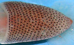
Figure. Ventral views of mantle photophores of H. meleagroteuthis. Left - Neotype, 48 mm ML, drawing extracted from Fig. 26a of Voss, 1969. Right - 26 mm ML, photographed aboard the R/V G. O. Sars, Mar-Eco cruise, central North Atlantic by R. Young.
- Compound photophores number 19-22 on margin of right eyelid.

Click on an image to view larger version & data in a new window
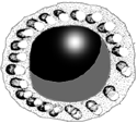
Figure. Lateral view of right eyelid of H. meleagroteuthis, neotype, 48 mm ML. Drawing from Voss, 1969 (Fig. 26h).
- Arms IV with 8-9 longitudinal series on arm base.

Click on an image to view larger version & data in a new window

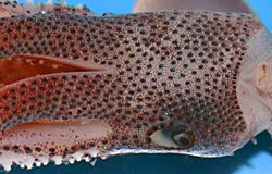
Figure. Ventral view of arms IV of H. meleagroteuthis. Top - Neotype, 48 mm ML. Drawing from Voss, 1969 (Fig. 26f). Bottom - 26 mm ML. Photographed aboard the R/V G. O. Sars, Mar-Eco cruise, central North Atlantic by R. Young.
- Fins
- Length 39-50% of ML; width 58-68% of ML.
- Spermatophores
- Length 1.6-2.1% of ML; sperm mass 28-39% of spermatophore length (SpL); cement body 15-37% of SpL; ejaculatory apparatus 28-36% of SpL and with single loop. Connective complex well developed.
- Hectocotylus
- Arms I in mature males elongated and modified in distal portions with sucker pedestals enlarged, producing a palisading effect to margin of arms; suckers in two series to tip.
Comments
The above information, except photographs, is taken from Voss (1969) and Voss, et al. (1998).
References
Voss, N. A. 1969. A monograph of the Cephalopoda of the North Atlantic: The family Histioteuthidae. Bull. Mar. Sci., 19: 713-867.
Voss, N.A., K. N. Nesis, P. G. Rodhouse. 1998. The cephalopod family Histioteuthidae (Oegopsida): Systematics, biology, and biogeography. Smithson. Contr. Zool., 586(2): 293-372.
About This Page
Drawings from Voss (1969) printed with the Permission of the Bulletin of Marine Science.

University of Hawaii, Honolulu, HI, USA

National Museum of Natural History, Washington, D. C. , USA
Page copyright © 2000 and
 Page: Tree of Life
Histioteuthis meleagroteuthis: Description continued
Authored by
Richard E. Young and Michael Vecchione.
The TEXT of this page is licensed under the
Creative Commons Attribution-NonCommercial License - Version 3.0. Note that images and other media
featured on this page are each governed by their own license, and they may or may not be available
for reuse. Click on an image or a media link to access the media data window, which provides the
relevant licensing information. For the general terms and conditions of ToL material reuse and
redistribution, please see the Tree of Life Copyright
Policies.
 Click on an image to view larger version & data in a new window
 Click on an image to view larger version & data in a new window

 Click on an image to view larger version & data in a new window


 Go to quick links
Go to quick search
Go to navigation for this section of the ToL site
Go to detailed links for the ToL site