Cephalopoda Glossary: Cirrate Male Reproductive Tract
Richard E. Young, Michael Vecchione and Katharina Mangold

Click on an image to view larger version & data in a new window

Figure. Two views of the male reproductive tract of Cirroctopus glacialis. Photographs taken by R. Young.

Click on an image to view larger version & data in a new window

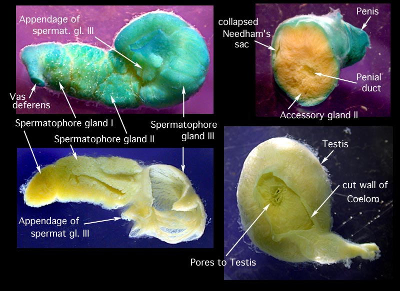
Figure. Views of different portions of the reproductive tract that have been cut open to show the different thicknesses of the glandular tissues. The spermatophore gland III in the lower left has had all spermatophores removed. The collasped portion of Needham's sac (upper right image) is not visible the the above photographs of the intact reproductive tract. The blue tint is due to staining with methylene blue. Photographs taken by R. Young.
About This Page

University of Hawaii, Honolulu, HI, USA

National Museum of Natural History, Washington, D. C. , USA
Katharina M. Mangold (1922-2003)

Laboratoire Arago, Banyuls-Sur-Mer, France
Page copyright © 2002 , , and Katharina M. Mangold (1922-2003)
 Page: Tree of Life
Cirrate Male Reproductive Tract Photographs
Authored by
Richard E. Young, Michael Vecchione, and Katharina M. Mangold (1922-2003).
The TEXT of this page is licensed under the
Creative Commons Attribution-NonCommercial License - Version 3.0. Note that images and other media
featured on this page are each governed by their own license, and they may or may not be available
for reuse. Click on an image or a media link to access the media data window, which provides the
relevant licensing information. For the general terms and conditions of ToL material reuse and
redistribution, please see the Tree of Life Copyright
Policies.


 Go to quick links
Go to quick search
Go to navigation for this section of the ToL site
Go to detailed links for the ToL site