Rondeletiola
Michael Vecchione and Richard E. Young


This tree diagram shows the relationships between several groups of organisms.
The root of the current tree connects the organisms featured in this tree to their containing group and the rest of the Tree of Life. The basal branching point in the tree represents the ancestor of the other groups in the tree. This ancestor diversified over time into several descendent subgroups, which are represented as internal nodes and terminal taxa to the right.

You can click on the root to travel down the Tree of Life all the way to the root of all Life, and you can click on the names of descendent subgroups to travel up the Tree of Life all the way to individual species.
For more information on ToL tree formatting, please see Interpreting the Tree or Classification. To learn more about phylogenetic trees, please visit our Phylogenetic Biology pages.
close boxIntroduction
Species of Rondeletiola are small, mostly less than 20 mm ML and can be common on mud and fine sand bottoms. Naef (1921-23) reported that about 5,000 were caught on one occasion in the Bay of Naples at a depth of 150-200 m.
Diagnosis
A sepiolin ...
- with a circular visceral photophore.
Characteristics
- Arms
- Arm sucker in two series.
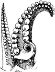
- Left arm I hectocotylized with 3 basal suckers proximal to copulatory apparatus with recurved lateral teeth; distal portion of arm not expanded.
- Arms II and III of male with enlarged suckers in ventral series.
- Tentacles
- Tentacular club with suckers in 8-16 series.
- Club suckers small, uniform in size.
- Photophores.
- Photophores fused into a circular organ in adults .
- Gladius
- Gladius absent.
Comments.
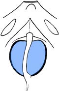
We place Inioteuthis capensis Voss (1962) into Rondeletiola based on what appears to be a large circular photophore embedded in the ink sac. In addition, the structure of the hectocotylus of this species appears to be closer to that of Rondeletiola than Inioteuthis.


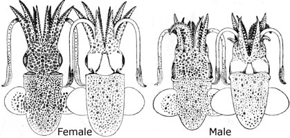
Figure. "Idioteuthis" capensis Voss , 1962. Left - Dorsal view of holotype, 10 mm ML. Middle - Ventral view of anterior mantle cavity with funnel cut open showing intestine and presumed photophore. Right - Oral view of hectocotylized left arm I.
Distribution
Mediterranean Sea and eastern Atlantic form Spain to the tip of South Africa, 15 m to upper slope.References
Naef, A. 1921-1923. Die Cephalopoden. Fauna e Flora del Golfo di Napoli, Monographie 35, Vol I, Parts I and II, Systematik, pp 1-863.
Voss, G.L. 1962. South African Cephalopods. Transactions of the Royal Society of South Africa, 36(4):245-272.
About This Page

National Museum of Natural History, Washington, D. C. , USA

University of Hawaii, Honolulu, HI, USA
Page copyright © 2004 and
 Page: Tree of Life
Rondeletiola .
Authored by
Michael Vecchione and Richard E. Young.
The TEXT of this page is licensed under the
Creative Commons Attribution-NonCommercial License - Version 3.0. Note that images and other media
featured on this page are each governed by their own license, and they may or may not be available
for reuse. Click on an image or a media link to access the media data window, which provides the
relevant licensing information. For the general terms and conditions of ToL material reuse and
redistribution, please see the Tree of Life Copyright
Policies.
- First online 02 September 2004
- Content changed 30 November 2003
Citing this page:
Vecchione, Michael and Richard E. Young. 2003. Rondeletiola . Version 30 November 2003 (under construction). http://tolweb.org/Rondeletiola/20040/2003.11.30 in The Tree of Life Web Project, http://tolweb.org/


 Go to quick links
Go to quick search
Go to navigation for this section of the ToL site
Go to detailed links for the ToL site