Enoploteuthis
Kotaro Tsuchiya


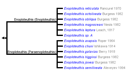
This tree diagram shows the relationships between several groups of organisms.
The root of the current tree connects the organisms featured in this tree to their containing group and the rest of the Tree of Life. The basal branching point in the tree represents the ancestor of the other groups in the tree. This ancestor diversified over time into several descendent subgroups, which are represented as internal nodes and terminal taxa to the right.

You can click on the root to travel down the Tree of Life all the way to the root of all Life, and you can click on the names of descendent subgroups to travel up the Tree of Life all the way to individual species.
For more information on ToL tree formatting, please see Interpreting the Tree or Classification. To learn more about phylogenetic trees, please visit our Phylogenetic Biology pages.
close boxIntroduction
Enoploteuthis contains the largest species in the family one, at least, can reach 130 mm ML. The species are most easily recognized by their larger tail compared to members of other genera in the family. The size of the tail is emphasized by the absence of fins along its sides. This contrasts with the narrow extension of the fins along the tails of members of related genera.
Brief diagnosis:
An enoploteuthid ...
- without large, black photophores at the tips of arms IV.
- with fins terminating well in advance of posterior tip of tail.
Characteristics
From Young, et al., 1998.- Arms
- Suckers present distally on Arms IV.
- Suckers present distally on Arms IV.
- Tentacles
- Manus of club with two series of hooks; marginal suckers absent.
- Manus of club with two series of hooks; marginal suckers absent.
- Buccal crown
- Typical chromatophores on aboral surface; may have light epithelial pigmentation on oral surface.
- Typical chromatophores on aboral surface; may have light epithelial pigmentation on oral surface.
- Photophores
- Tips of arms IV lack enlarged photophores.
- Nine to ten photophores on eyeball.
- Complex photophores of integument, in life, without red-colored filters.
- Fins
- Fins terminate well in advance of posterior tip of tail. (see title photograph).
- Fins terminate well in advance of posterior tip of tail. (see title photograph).
- Spermatangia receptacles
- Present at posterior junction of funnel-retractor muscles and head-retractor muscles.
- Present at posterior junction of funnel-retractor muscles and head-retractor muscles.
Comments
Enoploteuthis has only two clearly defined types of integumental photophores in contrast to other enoploteuthid genera which have three types of photophores on the ventral surfaces of the head, funnel and mantle. The most complex type is found in all species examined and seems to be distinctive of the genus.


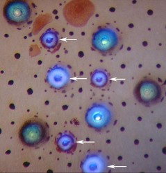
Figure. Integumental photophores of Enoploteuthis sp., Hawaiian waters. The arrows indicate "simple" photophores, of two sizes, with dark blue centers and a narrow white ring. The remaining five, slightly larger photophores are "complex" filtered photophores that exhibit three different physiological states. The central core in these shows varying degrees of transparency. The bright spot in the core of the most transparent photophore is a reflection off the axial color filter that lies buried in the center of the organ. The small dark spots and reddish patches are chromatophores.


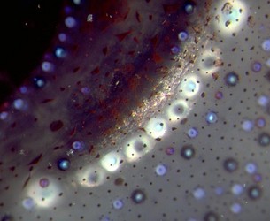
Figure. Ventral view of the ocular photophores (large white organs), beneath the smaller integumental photophores of Enoploteuthis sp, Hawaiian waters. The terminal ocular photophores are larger than the photophores between them. Photophores in the latter group alternate between having light and dark cores. The reason for this is unknown.
Classification
This genus consists of two natural groups: subgenus Enoploteuthis, which is characterized by having short, thin tentacles with small, thin clubs and subgenus Paraenoploteuthis, which has relatively long, thick tentacles with robust clubs (Tsuchiya and Okutani, 1988). The difference in the appearance of the tentacle is striking. Details of the club differ as follows:
| Tentacular club | Enoploteuthis (Enoploteuthis) | Enoploteuthis (Paraenoploteuthis) |
| Carpal locking-apparatus | Very elongate | Nearly circular |
| Relative hook size between the two series on manus | Often subequal | Very unequal |
| Dactylus suckers | 2 series | 4 series |
| Ventral protective membrane ("flap") on manus | Absent | Present |
| Two Crest ridges below Hood on upper beak | Absent | Present |


Figure. Oral views, tentacular clubs. Top - Club with characters of the E. (Enoploteuthis) group. Drawing from Tsuchiya and Okutani (1988). Note that the manus is narrower than the tentacle stalk. Bottom - Club with characters of the E. (Paraenoploteuthis) group. Drawing from Roper (1964). Note that the manus is distinctly broader than the tentacle stalk.
References
Burgess, L. A. 1982. Four new species of squid (Oegopsida: Enoploteuthis) from the Central Pacific and a description of adult Enoploteuthis reticulata. Fish. Bull. 80: 703-734.
Roper, C.F.E.1964. Enoploteuthis anapsis, a new species of enoploteuthid squid (Cephalopoda: Oegopsida) from the Atlantic Ocean. Bulletin of Marine Science of the Gulf and Caribbean, 14(1):140-148.
Tsuchiya, K. and T. Okutani. 1988. Subgenera of Enoploteuthis, Abralia and Abraliopsis of the squid family Enoploteuthidae (Cephalopoda, Oegopsida). Bulletin of the National Science Museum, Tokyo (series A) 14: 119-136.
Young, R. E., L. A. Burgess, C. F. E. Roper, M. J. Sweeney and S. J. Stephen. 1998. Classification of the Enoploteuthidae, Pyroteuthidae and Ancistrocheiridae. Smithsonian Contr. to Zoology, 586: 239-255.
About This Page

Tokyo University of Fisheries, Tokyo, Japan
Page copyright © 2018
All Rights Reserved.
- Content changed 11 October 2015
Citing this page:
Tsuchiya, Kotaro. 2015. Enoploteuthis . Version 11 October 2015 (under construction). http://tolweb.org/Enoploteuthis/19641/2015.10.11 in The Tree of Life Web Project, http://tolweb.org/


 Go to quick links
Go to quick search
Go to navigation for this section of the ToL site
Go to detailed links for the ToL site